
Early detection of the cancer-prone area in MRI scan is of great importance for the successful diagnosis and treatment of bone cancer. This paper proposes an approach to detect bone cancer in MR images using medical image processing techniques. A proposed approach has some pre-processing techniques which use Gabor filter to smoothen the image and remove the noise from an image. The segmentation is carried out by using super pixel segmentation and multilevel segmentation. This methodology is used for identifying the bone cancer by various pre-processing techniques like filtering and gray conversion. After filtering, edge detection and morphological operations are applied. In the second stage, super pixel segmentation is performed and some of the important features are extracted from the images. Then the extracted features are used to identify the bone cancer. In this project an approach of tumour detection using machine learning have been discussed and the data set for the performance analysis is MRI images.
Bone is the supporting skeleton of body and is hollow. The outer part of bones is a arrangement of tough tissue called matrix against calcium salts are laid down. The hard out layer is made with cortical bone, it covers trabecular bone inside, outside of bone covered with periosteum. Some bones are hallow and space is called medullary cavity which contains the soft tissue called bone marrow.
Endosteum is act as a tissue lining. At each end of the bone is a region of a softer shape of bone-like tissue called cartilage, it is softer than bone thatis made of fibrous tissue matrix assorted with a gel-like stuff that does not enclose much calcium. Most bones get going out as cartilage. The body then put downs calcium onto the cartilage to form bone. After the bone formation, some cartilage may stay at the ends to act as a bolster between bones. This cartilage, along with ligaments and some other tissues join bones to form a joint. Bone itself is very stiff and muscular. Bone is able to hold up as much as 12,000 pounds per square inch.
It takes as much as 1,200 to 1,800 pounds of pressure to break the thigh bone. The bone contains 2 kinds of cells. The osteoclast is the cell thatform new bone, and the osteoclast is the cellthat softens old bone. some bones the marrow is greasy tissue. The marrow in other bones is a concoction of fat cells and blood-forming cells. The blood-forming cells fabricate red blood cells, white blood cells, and blood platelets.Other cells in the marrow include plasma cells, fibroblasts, and reticuloendothelial cells
Bone refashion activity is only due to Cancer cells in the Bone. Normal bone is indefatigably being amended, or conked out and rebuilt. Cancer cells offend the balance for growth and formation of cell in bone. If cancer cells are in the bones, then the structure of bone is bent at a higher rate when compared to normal bone rate. Mostly bone cancer will be of primary or secondary . Primary bone cancer occurs in the bone. Whereas secondary bone cancer happens anywhere in the Body.
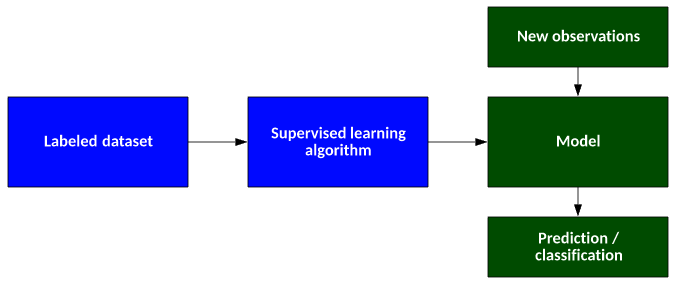
Machine learning covers these areas:
1.Classification assign a category to each object (OCR, text classification, speech recognition).
2.Regression predict a real value for each object (prices, stock values, economic variables, ratings).
3.Clustering partition data into homogeneous groups (analysis of very large data sets).
4.Ranking order objects according to some criterion (relevant web pages returned by a search
engine).
In this project a method is introduced to detect bone cancer by using machine learning algorithm. The main objective is to detect the tumor present in the bone, but most of the times happens that in methods of tumor detection the images obtained comes up with the greater noise factor which restrict the area to operate as it doesn’t give the exact location of tumor and the affected tissues. Hence in this paper a novel approach have been proposed which will comprised ofthe number of stageswhichwill ultimately lead to the proper detection of enchondroma tumor i.e. bone tumor.
A simple flow chart for the proposed system as follows:

Tumor Identification
The bone tumor is identified by simply calculating the mean pixel intensity of segmented image.
Mathematically, the mean pixel intensity can be calculated as:

Tumor Detection
After the tumor identification process itis last step to detect the tumor which can be carried out by using the MATLAB function for connected components which will simply select out the area with maximum connected component and the remaining area will be discarded.

The proposed system of bone tumor detection with superpixel segmentation is implemented using python. Also the detection of brain cancer is carried out with the given set of images. The proposed system is specially dedicated for brain tumor detection . The same system can be further extended to identifying the stages of cancer.